Ligadura de arteria carótida común y exsanguinación como modelo experimental de isquemia cerebral focal en ratas
DOI:
https://doi.org/10.33017/RevECIPeru2013.0017/Keywords:
Focal cerebral ischemia, ligation of left common carotid arter, ex sanguinationAbstract
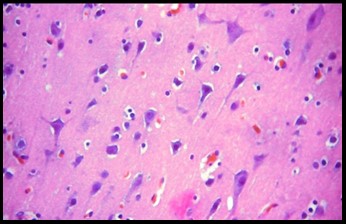
Cerebral ischemia is the pathophysiological process characterized by dysfunction of a portion of brain tissue secondary to decreased flow in specific brain artery. The best tool available today to study the pathophysiology of cerebral ischemia are experimental models, which allow a simple way to address the care of the condition which is characterized by complexity and heterogeneity. Whereas research on this field is unlimited, not only because of the importance but by the social cost of the disease, the study of new experimental methodologies to achieve better tools for scientific rigor in pathophysiological knowledge, treatment and prevention of cerebral ischemic disease. So we set out to determine whether the common carotid artery ligation and ex sanguination left can be used as a model of focal cerebral ischemia in rats. We used 36 male rats of 8 to 9 weeks of age, with 200 ± 20g of weight, were maintained in standard lighting conditions, daily cycles of 12 hours of light and 12 of darkness were used at an ambient temperature of 24-25 ° C, the specimens were divided into two groups: Sham Groups and Experimental Group (operated). We proceeded to the bleeding via cardiac puncture, and extracted from each rat 10% of circulating blood volume. Under aseptic and antiseptic conditions, an incision was made in the midline of the neck and dissected the skin, subcutaneous tissue and muscle to be able to identify the left common carotid artery (CCA) and ligated with 5-0 nylon suture to interrupt circulation way of ipsilateral blood, the wound was sutured and received lidocaine finally allowed to rest the animal at a slightly higher environmental chambers previously made temperature. After 24 hours of surgery applied, the specimens were sacrificed by overdose of anesthetic and removal of the brain and histopathological analysis of coronal sections in three regions neocortex, hippocampus and basal ganglia was performed, being the most vulnerable areas to damage ischemic injury. It was found that the major characteristics that indicate neuronal cell damage demonstrated in the experimental group with p <0.01 and the area with the highest incidence of injury is the basal ganglia, p <0.01. It was concluded that ligation of left common carotid artery and ex sanguinación produces localized neuronal damage and the most affected area is the basal ganglia.


